Concept explainers
To review:
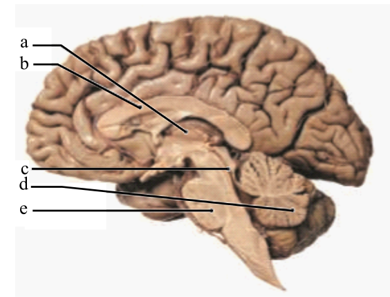
Label the following terms in the given diagram of the midsagittal section of the brain: pons, corpus callosum, cerebellum, thalamus, and cerebral aqueduct.
Introduction:
The brain is the prime organ of the CNS (central nervous system). It is divided into six major regions on the basis of structure and function, which are cerebrum, cerebellum, medulla oblongata, pons, diencephalon, and mesencephalon.
Explanation of Solution
The structures of the midsagittal section of the brain are labeled as below:
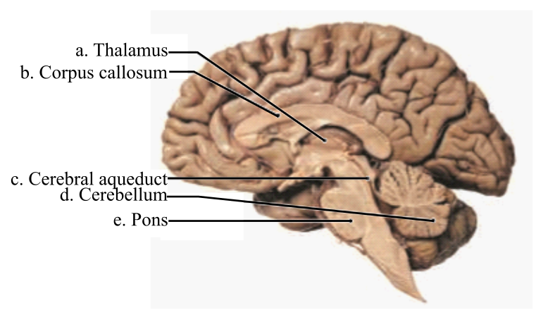
Thalamus: It is a part of the diencephalon that is further divided into right and left thalamus. This region is assigned the duty to relay and process sensory information across the brain.
Corpus callosum: It is a bundle of commissural fibers that connects the two cerebral hemispheres. It functions to transfer information from one hemisphere to another. It contains the largest portion of the white matter in the brain.
Cerebral aqueduct: It is a part of the mesencephalon or midbrain. It connects the third and fourth ventricles within the diencephalon and mesencephalon respectively. It is filled with cerebrospinal fluid (CSF), which flows into the ventricles.
Cerebellum: It is a part of the hindbrain and called as the small brain. It functions to relay motor sensations and is involved in intellectual development. It also maintains the posture of the body.
Pons: It is a part of the brainstem and is located below the midbrain, above the medulla oblongata and anterior to the cerebellum. It functions to relay sensory information between the thalamus and the cerebellum. It is responsible for the maintenance of body posture, hearing, taste, touch, pain, facial expression, chewing, salivation, and secretion of tears.
Therefore, the structures of a midsagittal section of the brain are:
| a | Thalamus |
| b | Corpus callosum |
| c | Cerebral aqueduct |
| d | Cerebellum |
| e | Pons |
Want to see more full solutions like this?


- Label all the nerve chords, thoracic gaglia and abdominal ganglia. State thr number of nerve chords , thoracic ganglia and abdominal ganglia isa present in the “picture”.arrow_forwardLabel the following numbers: 50, 51, 53, 55, 56, 59, 60, and 62 of the cerebrum in lateral view (A) and diencephalon in sagittal view (B)arrow_forwardFor each of the following brain structures, write G (for gray matter) or W (for white matter) as appropriate.__(1) cortex of cerebellum__(2) pyramids__(3) internal capsule and corona radiata__(4) red nucleus__(5) medial lemniscus__(6) cranial nerve nuclei__(7) cerebellar pedunclearrow_forward
- Match the names of the cranial nerves in column B to the appropriate description in column A.arrow_forwardName 12 cranial nerves and list their function.arrow_forwardMatch each cranial nerve with the type of information it carries (sensory, motor, or mixed). CN I (olfactory nerve) CN II (optic nerve) CN III (oculomotor nerve) CN IV (trochlear nerve) CN V (trigeminal nerve) CN VI (abducens nerve) CN VII (facial nerve) CN VIII (vestibulocochlear nerve) CN IX (glossopharyngeal nerve) CN X (vagus nerve) CN XI (spinal accessory nerve) CN XII (hypoglossal nerve)arrow_forward
- A 40 year old male has just been diagnosed with amyotrophic lateral sclerosis, better known as Lou Gehrig’s Disease. A recent MRI has shown significant demyelination of the gracile nucleus of the dorsal columnar-medial lemniscal pathway. Describe the physiology of this pathway and discuss what neurological deficits this patient will experience due to his lesion.arrow_forwardMatch each cranial nerve with the type of information it carries (sensory, motor, or mixed). Study your notes first, and try to do this without looking. Group of answer choices (sensory, mixed, motor) -CN I (olfactory nerve) -CN II (optic nerve) -CN III (oculomotor nerve) -CN IV (trochlear nerve) -CN V (trigeminal nerve) -CN VI (abducens nerve) -CN VII (facial nerve) -CN VIII (vestibulocochlear nerve) -CN IX (glossopharyngeal nerve)…arrow_forwardMatch the names of the cells in column B with the function they perform, shown in column A.Column A Column B(1) line the central cavity of the brain (a) astrocytes(2) form myelin in the CNS (b) ependymal cells(3) form myelin in the PNS (c) microglial cells(4) remove neurotransmitters in the CNS (d) oligodendrocytes(5) regulate ionic composition of thefluid around neurons in the CNS (e) satellite cells (f) Schwann cells(6) CNS phagocytesarrow_forward
 Human Physiology: From Cells to Systems (MindTap ...BiologyISBN:9781285866932Author:Lauralee SherwoodPublisher:Cengage Learning